How do we diagnose cancer in pets? - Tests and diagnostics
When a dog or cat shows a lump, mass, or other suspicious symptoms, it is important to quickly and carefully investigate whether cancer is present. A cancer diagnosis is made through a combination of physical examination, imaging techniques, and tissue analysis.
In this article, we discuss how cancer is diagnosed in pets and which methods veterinarians use to arrive at a definitive diagnosis.
Physical examination
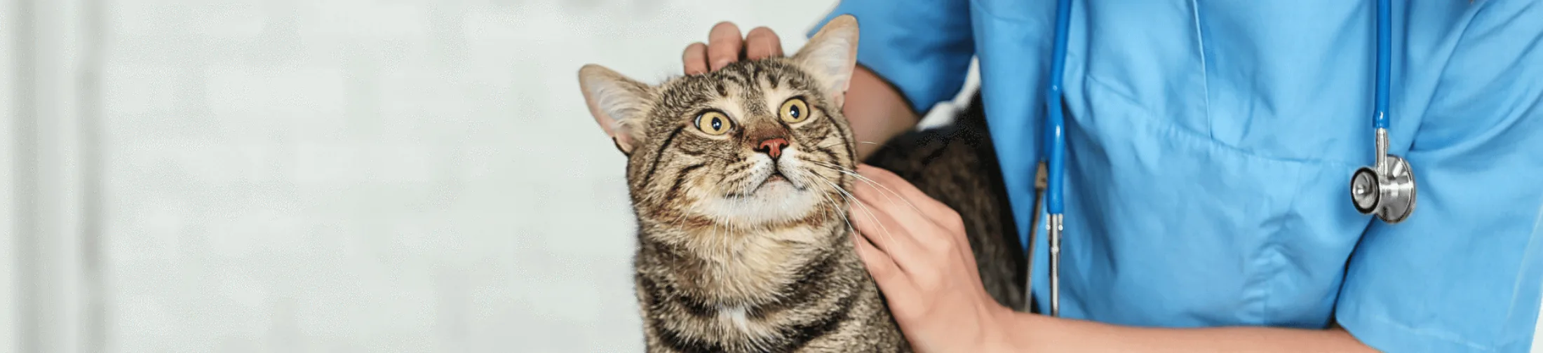
The first step is a thorough physical examination. The veterinarian feels for lumps, swellings, and enlarged lymph nodes, listens to the heart and lungs, and assesses the animal's overall condition. Although a physical examination can provide clues, it is never sufficient on its own to definitively diagnose cancer.
Imaging
Imaging allows veterinarians to look inside the body at organs and tissues. This helps detect tumors, assess their size and location, and check for metastases.
Ultrasound
Ultrasound uses sound waves to create images of organs. This is especially useful for assessing the liver, spleen, kidneys, lymph nodes, and abdominal cavity. In addition, ultrasound can help guide targeted biopsies or aspirations of organs or fluid, allowing for a more accurate diagnosis.
X-rays (radiographs)
X-rays are often used to assess the chest and bones. They are valuable for detecting lung metastases and for diagnosing bone cancer.
CT scan
A CT scan provides detailed cross-sectional images of the body. It is often used for tumors in the head, chest, and abdomen, and is very useful for visualizing the exact extent of a tumor. A CT scan is also a valuable tool in planning surgical procedures, and with contrast medium it can help better detect possible metastases.
MRI scan
An MRI scan mainly provides a great deal of information about soft tissue and the nervous system. It is often used for brain tumors or tumors of the spinal column.
Additional imaging techniques
In addition to MRI and CT, other techniques can also be used to better detect metastases, such as the use of methylene blue or ICG fluorescence. These dyes make lymph nodes visible and help locate possible metastases.
Biopsy and aspiration
A definitive cancer diagnosis is always made by examining cells or tissue. This can be done in two ways:
Fine needle aspiration
A thin needle is used to draw cells from a lump or lymph node. These are examined under a microscope (cytology). It is a quick and usually minimally invasive method, but sometimes not sufficient for a definitive diagnosis.
Biopsy
In a biopsy, a piece of tissue is surgically removed for histological examination. This provides much more information about the nature of the tumor and is usually necessary to determine a treatment plan.
Why is tissue examination so important?
Only histological or cytological examination can determine with certainty whether a tumor is benign or malignant. It also provides information about the tumor's aggressiveness and helps determine the best treatment.
Prognosis and next steps
After the diagnosis is made, the veterinarian discusses treatment options with the owner. This can range from surgery to chemotherapy, radiation therapy, or palliative care, depending on the type and stage of the cancer.
Want to know more?
Read more about oncology and the various treatment options available at Nesto. Our specialized veterinarians combine modern diagnostics with extensive experience to provide the best possible care for your pet.
Frequently asked questions (FAQ)
How can I recognize cancer in my pet?
Lumps, lethargy, weight loss, reduced appetite, and changes in behavior can all be signs.
Is a physical examination enough to diagnose cancer?
No, additional tissue examination is always needed for a definitive diagnosis.
What is the difference between a fine needle aspiration and a biopsy?
A fine needle aspiration collects individual cells, while a biopsy provides a piece of tissue that gives more detailed information.
Why is imaging important?
To determine the size, location, and any possible metastases of a tumor.
Is every lump examined?
Yes, every lump in a dog or cat deserves attention and should be assessed by a veterinarian.

Nesto
Share on