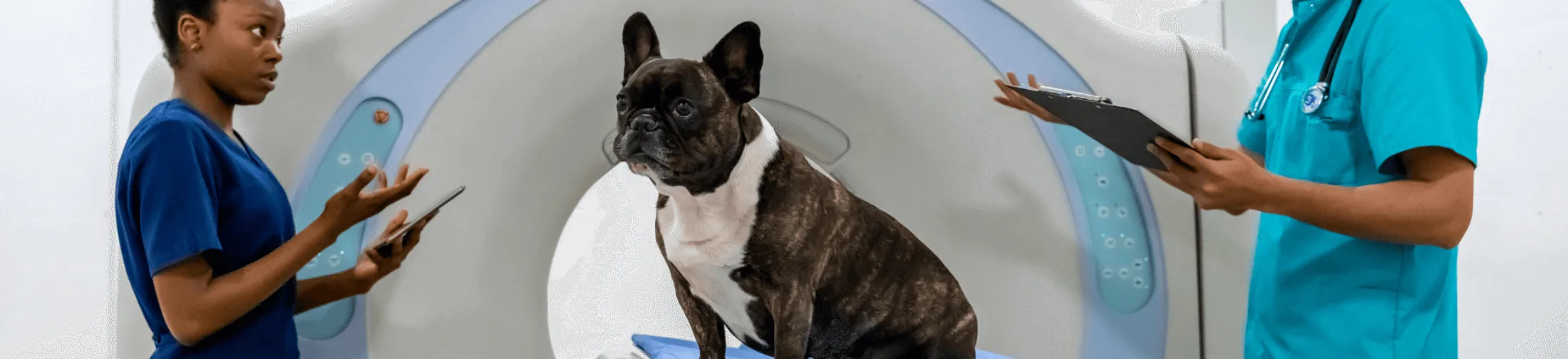
MRI and CT scans in pets - Neurological examination
MRI (magnetic resonance imaging) and CT (computed tomography) are advanced imaging techniques that play a crucial role in diagnosing neurological conditions in dogs and cats. These examinations make it possible to view the brain, spinal cord, and spine in detail.
In this article, we explain how MRI and CT work, in which situations they are used, and what value the results have for your pet's treatment.
What is an MRI and CT scan?
MRI uses magnetic fields and radio waves to create detailed images of soft tissue, such as the brain and spinal cord. CT, on the other hand, uses X-rays to reconstruct cross-sections of the body and is particularly suited to bone structures and acute injuries.
When is MRI or CT used?
Both techniques are often used for neurological complaints such as:
- Epilepsy and other seizures
- Paralysis or coordination disorders
- Back pain or neck pain
- Trauma to the head or spine
- Inflammatory or infectious diseases of the nervous system
- Balance disorders
Preparing the animal
Because MRI and CT require the animal to lie completely still, dogs and cats are always examined under general anesthesia. Prior to the scan, a pre-anesthetic examination is carried out, including a physical check and, if necessary, blood tests, to minimize risks.
How does the examination work?
For MRI, the animal is placed inside a strong magnet and images are created using radio waves. This usually takes 30 to 90 minutes. CT scans are faster (a few minutes) and use X-rays. Contrast fluid is sometimes administered to make abnormalities more visible.
What do MRI and CT show?
MRI is particularly well suited to detecting abnormalities in soft tissue such as the brain, spinal cord, and nerves. CT is better at assessing bone structures such as the skull and vertebrae, but can also reveal tumors and hemorrhages. The choice between MRI and CT depends on the suspected condition and the area to be examined.
Recovery after the examination
After the scan, the animal wakes up calmly from anesthesia. Most animals recover quickly and can go home the same day. Some animals may experience mild drowsiness or nausea due to the anesthesia, but this usually resolves within 24 hours.
Safety of MRI and CT
MRI does not use radiation and is completely safe, provided there are no metal implants present. CT does use X-ray radiation, but in controlled amounts that do not cause harm with occasional use.
Want to know more?
Read more about our services in neurology and imaging. Our specialized veterinarians at Nesto guide you and your pet through the entire process of MRI and CT diagnostics and the subsequent treatment.
Frequently asked questions (FAQ)
What is the difference between MRI and CT?
MRI is better for soft tissue such as the brain, while CT is better for bone structures and acute injuries.
Does my pet need anesthesia?
Yes, full anesthesia is needed for both MRI and CT to prevent movement.
When is MRI chosen over CT?
MRI is often chosen when a brain or spinal cord condition is suspected, while CT is used for bone injuries.
Is MRI or CT dangerous for my pet?
MRI is completely safe; CT uses radiation, but in safe amounts.
How quickly are the results available?
The images are produced immediately, but interpretation by the specialist usually follows the next day, or slightly later depending on workload.

Nesto
Share on